Caso del mese:
Infantile acute hemorrhagic edema and Rotavirus infection
Dott.ssa
M.Lombardi, Dott. G. Lo Scocco
Divisione Dermatologia Ospedale S.Maria Nuova
Reggio Emilia
Infantile acute hemorrhagic edema (AHE) of the skin is a distinctive cutaneous disorder characterized by large rosette-shaped purpuric lesions on the face and limbs, and by acral edema accompanied by fever occurring, almost exclusively in children between the ages of 4 months and 2 years, during winter. Spontaneous resolution normally follows within 3 weeks. The nosologic position regarding the disease is still debated. Although some have suggested considering AHE a purely cutaneous variant of purpura of Schönlein Henoch (HSP), most Authors prefer to regard it as a separate clinical entity among cutaneous small vessel vasculitis of childhood.
We describe a child with AHE with a concurrent rotavirus infection which has not been reported in association with AHE.
Case report
An 11-month-old girl was seen for purpuric skin lesions occurring the day previous to her visit. Her birth weight was 3805 gr and her length 52 cm. There was no history of recent infectious diseases or drug therapy.
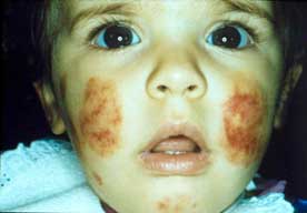
The cutaneous examination showed many symmetrically distributed, round or oval, ecchymotic, purpuric, targetoid plaques ranging from 2 cm to 4 cm in diameter, localized on the face ( Figure 1), especially cheeks and ears, and extremities, in particular lower limbs. Some plaques showed a cockade pattern, while others coalesced to form large purpuric lesions with polycyclic borders. The ears appeared oedematous (Figure 2); petechiae on the gums and haemorrhagic lachrymation were also noted. The remainder of the physical examination was not noteworthy and there were no signs of systemic involvement. During the following six days the patient had a mild fever and several diarrhoeic episodes. No other systemic symptoms were noted
.
Laboratory studies revealed: hematocrit 39%, haemoglobin 13,2 mg/dl, leucocytes 9,500/mm3 (55% neutrophils, 38% lymphocytes, 5% monocytes, 0% eosinophils), platelet count 403000/ mm3, erythrocyte sedimentation rate 26 mm/hour, C3 120,7 mg/dl (normal 60-158mg/dl), C4 48,9 mg/dl (normal 18-105 mg7dl), CPR 1,07 mg/dl (normal 0,00-0,5 mg/dl) , fibrinogen 477mg/dl (normal range 150-450mg/dl). Quantitative serum immunoglobulins were as follow: IgG 519,5 mg/dl (normal range 241-613 mg/dl), IgA 67,5 mg/dl (normal range 10-46 mg/dl), and IgM 109 mg/dl (normal range 26-60 mg/dl); in the serum electrophoresis α2 globulin and β globulin were slightly increased, while γ globulin reduced; urea, electrolytes and liver function tests were within normal limits. The urine analysis was normal, with the presence of rare leucocytes in the sediment. Tests for ANA and anti-DNA were negative, as well as a tuberculin skin test (5U).
Nasopharyngeal cultures for Streptococcus pyogenes, serologic tests for Salmonellae, Shighellae, TORCH, Yersinia enterocolitica, Campylobacter jejuni, Rickettsia conori, Mycoplasma pneumoniae and Borrelia burgdorferi were all negative. Stool sample cultures grew Rotavirus. A first stool sample was positive for occult blood, a second one slightly positive and a third one, after one week, definitely negative. Parents denied permission for skin biopsy. The skin lesions, as well as fever and diarrhoea, resolved without treatment within two weeks. No relapse was observed after a two year follow-up. Repeated stool sample cultures gave negative results. A retrospective diagnosis of AHE was made.
Discussion
The clinical features of our case, characterized by a dramatic, acute onset of typical large, symmetrical, annular purpuric plaques on the face, ears and limbs, which showed a spontaneous recovery within two weeks, were consistent with the diagnosis of AHE. Routine laboratory tests of patients with AHE are not diagnostic, disclosing normal results, or, as in our patient, elevated erythrocyte sedimentation rate and increase of α2 globulin.
A clinical disorder corresponding to AHE was firstly described in 1913 by Snow et al. (1) who reported a patient as “ Purpura, urticaria and angioneurotic oedema of the hands and feet in a nursing baby”. Afterwards, many cases of this condition have been reported in the European literature under different clinical terms, among which Finkelstein’s disease (2), Seidlmayer's “cockade” purpura or syndrome (3). AHE has rarely been reported in the United States, probably due to cases of AHE reported as SHP of infancy (4,5). Until now, approximately 80 cases of AHE have been reported in the literature, although the disease could be underreported (4,6,7).
AHE is considered an uncommon form of cutaneous leukocytoclastic vasculitis which occurs in infants younger than 2 years, the mean being 12,4 months (7-9). Its aetiology remains unknown, although a history of recent upper respiratory or urinary tract infection treated with antibiotics or immunizations is found in 75% of patients (8-10). Thus, AHE is considered to represent an immune-complex mediated disease. The most reported infective agents include Staphylococci, Streptococci and among viruses, Adenovirus, although many other agents, such as Escherichia coli, or Mycobacteria, have been reported in association with cases of AHE. Diarrhea has been observed during episodes of AHE and related to Coxsackievirus or Campylobacter Mucosalis in stools (10-11). Interestingly, our 11-month old patient presented gastrointestinal symptoms during the skin eruption related to a Rotavirus infection, which has been reported in association with various cutaneous manifestations such as Gianotti-Crosti syndrome or exanthems (12-13), but is not known as a cause of AHE. Rotavirus infections are important aetiological agents of nosocomial infections in childhood, and are the main pathogens of infantile diarrhea in winter months. Thus, it is difficult to discern whether the infection was the primary cause that triggered the disease. Culture of stool-samples are advisable in laboratory investigations of AHE to confirm the possible aetiological role of Rotavirus.
A history of drug intake before the onset of the cutaneous eruption is present in many cases of AHE. The drugs include various antibiotics and anti-inflammatory drugs (7,10,14).
Histologic features of AIHO are consistent with small-vessel vasculitis of both capillaries and post capillary venules of the upper and the middle dermis, showing in the dermis typical leukocytoclastic vasculitis with or without fibrinoid necrosis and a deep perivascular and interstitial infiltrate composed mostly of neutrophils with abundant nuclear dust (7,15). Results of direct immunofluorescence study are not significant for the diagnosis, for the possible presence of IgM, C3 and fibrinogen; in addition IgA deposits can be found in one third of the cases, while C1q and IgG have been occasionally observed (7,10). Since histological findings of AHE are common to other forms of leukocytoclastic vasculitis (16), in particular HSP and/or vasculitis related to drug hypersensitivity and viral hypersensitivity, a clinicopathologivc correlation is needed for the diagnosis .
Other conditions that must to be differentiated are Sweet’s syndrome, erythema multiforme, Kawasaky disease, purpura fulminans, trauma-induce purpura and granuloma faciale. Differential diagnosis is usually possible on the basis of history, physical examination, laboratory investigation and histologic examination.
It is still debated whether AHE should be regarded as a separate clinical entity within the spectrum of leukocytoclastic vasculitis or just a benign variant of HSP. Amitai et al. suggested that AHE cannot be considered a distinctive syndrome, but a benign variant of SHP pointing out a similar pathogenesis of both diseases, but a different distribution of purpuric lesions and with predilection of the face in infants (AHE) and buttocks and lower extremities in older children (HSP). In addition, they postulated that the proportionally larger head and face in infant with a corresponding increase in blood supply would render them more susceptible to facial purpura (4). Cases of AHE and HSP overlap have been reported (16); however, most Authors consider these two to be distinct clinical and pathological entities with some distinctive characteristics (7,9,10): AHE is observed before 2 years of age and is confined to the skin, with less polymorphic skin lesions with respect to HSP, a more benign course without relapses, and IgA deposits on immunofluorescence studies in only one third of the cases. On the other hand, HSP has an age peak between 3-7 years, papulopetechial or urticarial lesions manifests on the extensor surfaces of the legs and buttocks, and shows frequent relapses. Renal, gastrointestinal, and joint involvement are common. The deposition of IgA in the basal membrane is its the immunopathological hallmark of HSP. It has been suggested that the immune complexes remain in the cutis in AHE, while they spread throughout skin, kidney and gastrointestinal tract in HSP.
References
1. Snow IM. Purpura, urticaria and angioneurotic edema of the hands and feet in a nursing baby. JAMA.1913; 61: 18-9
2. Long D, Helm F. Acute hemorragic edema of infancy: Finkelstein’s disease. Cutis 1998; 61:183-4
3. Cox NH. Seidlmayer's syndrome: postinfectious cockade purpura of early childhood. J Am Acad Dermatol 1992 ;26:275
4. Amitai Y, Gillis D, Wasserman D, Kockman RH. Henoch Shöenlein purpura in infants. Pediatrics 1993; 92: 865-867
5. Allen DM, Diamond LK, Howell DA. Anaphylactoid purpura in children (Schöenlein-Henoch syndrome). Am J Dis Child 1960; 99: 833-54
6. Crowe MA, Jonas PP. Acute hemorragic edema of infancy. Cutis 1998; 62: 65-6
7. Saraclar Y, Tinaztepe K, Gonul A, Tuncer A. Acute hemorrhagic edema of infancy (AHEI) -a variant of Henoch-Schönlein purpura or a distinct clinical entity ? J Allergy Clin Immunol 1990; 86: 473-483
8. Ince E, Mumcu Y, Suskan E, et al Infantile acute hemorrhagic edema: a variant of leucocytoclastic vasculitis. Pediatr Dermatol 1995; 12: 224-7
9. Pride HB, Maroon M, Tyler W. Ecchymoses and edema in a 4-month old boy. Pediatr Dermatol 1995: 4:373-5
10. Legrain V, Lejean S, Taieb A, et al. Infantile acute hemorrhagic edema of the skin: studies of ten cases. J Am Acad Dermatol 1991; 24: 17-22
11. Gonggrypp LA, Todd G. Acute hemorragic edema of chidhood (AHE). Pediatr Dermatol 1998; 15:91-6
12. Di Lernia V. Gianotti-Crosti sindrome related to rotavirus infection. Pediatr Dermatol 1998; 15:485-6
13. Ruzicka T, Rosendahl C, Braun-Falco O. A probabile cause of rotavirus exanthem. Arch Dermatol 1985; 253-4
14. Gelmetti C, Barbagallo C, Cerri D et al. Acute hemorragic oedema of the skin in infants: clinical and pathogenetic observations in seven cases. Pediatr Dermatol News 1985; 4:23-34
15. Cunningham BB, William A.C, Lynne R.E. Neonatal acute hemorrhagic edema of chilhood: a case report and review of the English literature. Pediatric Dermatology 1996; 13: 39-44
Figure legends:
Figure 1. Round or oval purpuric targetoid plaques on the face.