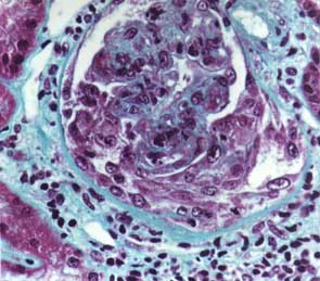
is also visible (Trichrome stain x 250).
|
|
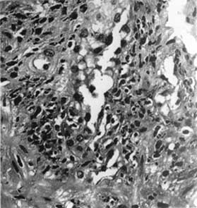
Fig.
1: Numerous polymorphonucleated cells
infiltrate the wall of a small artery in the submucosa of the ileum (Ematoxylin
eosin x 400). |
|
|
Fig.
2: Glomerular cell proliferation with
crescent formation, fibrin deposition and some polymorphs. A pericapsular
inflammatory infiltrate is also visible (Trichrome stain x 250). |
|
|
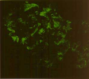
Fig.
3: Heavy deposition of IgA in the mesangium
and, to a lesser extent, in the capillary walls (Immunofluorescence x
250). |