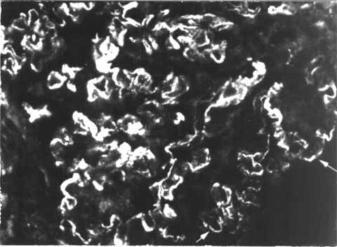
Figure
2: Immunostaining
of glomerular capillaries with fluoresceinated anti-IgG
antibody which is
localized in small granules along the basement membrane (arrows)
(x 400).
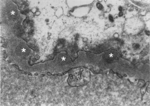
Figure
3: Electron
micrograph showing typical electron dense deposits (asterisks)
on the
epithelial side of the capillary wall. The foot process organization is
completely
lost and the podocyte cytoplasm contains numerous aggregates of
microfilaments
(x15,000, uranyl acetate and lead citrate staining).